Overview
The development of new drugs and estimations of toxicity in compounds used in common products like cosmetics require models imitating conditions close to in vivo status. The usage of human cell lines offers a great possibility to create models mimicing the cellular environment in human body. Working with such models, the effects of subtances can be tested very fast due to relative short cultivation times of cells. With genetic modifications the creation of new biosensors are possible which enable versatile possibilities for determination of the cellular status. The work with such methods and other possibilities in cell culture is done by our group which can be found in this page.
Cell Lines and Methods
Our working group gained over the last years great expertise in the field of cell culture and biosensor development. Today we are working with four following cell lines: HaCaT (skin cells), A549 (lung cells from an adenocarcimoa), HepG2 (liver cells from an hepatocellular carcinoma) and MMC (cell line created by clinic of Saalfeld). Available assays for cell culture are the MTT-assay as well as the resacurin-assay which we optimized for absorption and fluorescence measurement. With our fluorescence microscop and cLSM we are able to perform fluorescence staining of cell compartiments or specific biomarker or to detect GFP expression of genetically modified cells. Our methods for cell culture assays include beside the usual 2D-cell-culturing also the possibility of 3D-cell-culture. We also gained great experience in primary cell culture including the isolation of tumor cells and the optimization of cell culturing. A more detailled description of our work can be found in the following passages.
3D Cell Culture
Though 2D cell culture offers great possibilities for determination of the effects of substances, it lacks regarding the mimic of cellular interaction with the surrounding matrix and other cells. To reduce the emergence of the problem, three-dimensional culture of cells is recommended. Since cells are closely connected in a three dimensional geometry, nutrient and waste transport as well as the transport of substances is changed dramatically imitating the conditions in vivo much better. This can lead to different results of substances effects very different to 2D-cell culture.
In our labratory we created 3D-cell culture with HepG2-cells using different substrates for gelation. Notably these substrates were gelatin, collagen and matrigel that were cross-linked via transglutaminase. 3D-culture using three different gels showed different activity of liver cells displayed by urea and albumin formation. The highest production of these components was achieved in three dimensional matrigel matrix. In this gel, cells were ecnapsulated and formed spheroids that could live for more than 16 days.
Cell-Based Biosensors
During the development of new substances or materials it is necessary to know if a contact with humans can lead to any harmful reactions. To get detailed information about any reactions, a model close to the real exposure situations is required. Such a model can be easily generated by the use of an appropiated cell line. To achieve a detailed a sensitive read-out of any possible reactions, the implementation of genetically modified stress-induced proteins with a marker is well suited. Working with such genetically modified cells, effects of substances in a molecular level can be detected in a qualitative and quantitative way.
In our group we developed several of such biosensors. One of our biosensor is based on the expression of heat shock proteins caused by oxidative stress. This cell-based sensor was developed for HaCaT cells to test skin irritation effects and the lung cell line A549 to test cytotoxic effects of nanoparticles exposed to the lung. To create this biosensor, cells were transfected with a reporter-promotor-construct that inserted modified heat shock proteins (HSP) coupled with GFP (Green Fluorescent Protein). If oxidative stress due to harmful substances occurs, the regulation of MAP-kinase-pathway begins which leads to an expression of different HSPs. Via fluorescence microscopy cells with GFP-coupled HSPs can be detected. For each cell line, a certain heat shock protein was applicable. In HaCaT cellsthe biosensor worked great with a modified HSP 70B. Unfortunately this HSP showed bad results in the cell line A549 while a modified HSP 72 was usable. Working with sublethal concentrations, qualitative and quantitative measurements of cytotoxic can be tested.
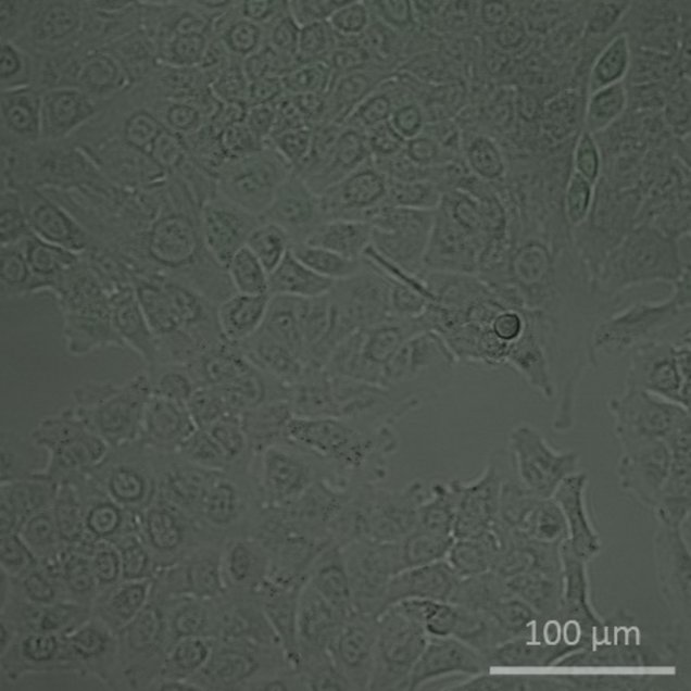
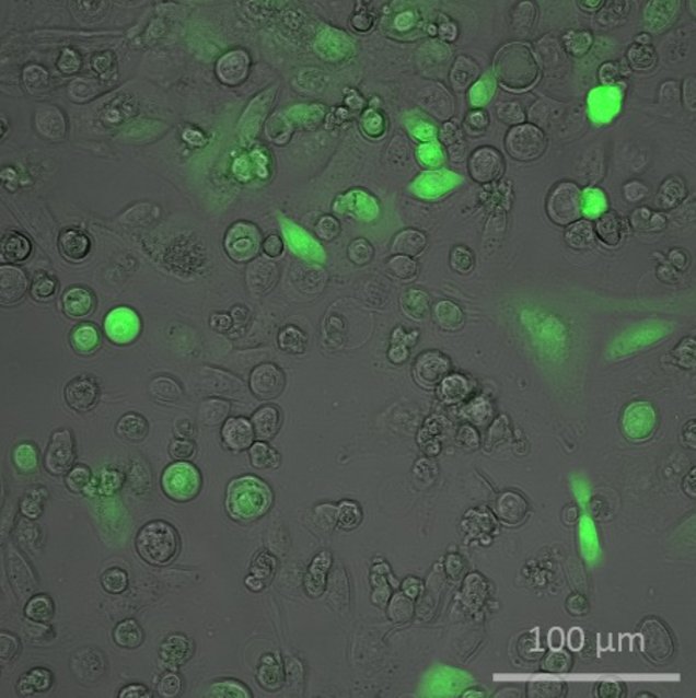
| A549 cells before incubation with 50 μm Cadmium Chloride nanoparticles | A549 cells 44 h after incubation with 50 μM Cadmium Chloride nanoparticles |
Another biosensor was developed for the effect of nanoparticles exposed to the liver displayed by HepG2 cells. For a better mimic of in vivo conditions of a real liver, a 3D cell culture was applied to these cells. To achieve 3D-structures, Matrigel was added in the medium and was cross-linked with addition of transglutaminase leading to cell spheroids.
The biosensor is based on the NF-κB-pathway which is regulated in the cytoplasm. Therefor a reporter-promotor-construct was transfected to the cells which caused the expression of alkaline phosphatase. If the NF-κB-pathway is activated, transcription factors are expressed which bind to the reporter-promotor construct. This will cause the expression of alkaline phosphatase which is secreted out of the cell. Adding the substrate SEAP to the media, a color change of the substrate will occur that causes a signal measurable at 405 nm. This signal is proportional to the toxicity of the nanoparticles and enables a concentration-based measurement of toxicity. The combination of 3D-cell-culture and biosensors shows a powerful tool to achieve investigations of nanoparticle cytotoxicity with conditions closer to in vivo.
Primary Cell Culture
Primary cell culture offers multiple advantages towards immortalized cell lines since there are no genetic alterations. This offers unique investigations with results that are closer to real organisms as for instance the test of chemosensitivity of tumors which is done by our group. For establishment of primary cell culture we used cryo und fresh lung cancer biopsies and isolate tumor cells from these samples. Therefor we investigated systematically several parameters that could affect the efficieny of the isolation starting from the disaggregation method up to data from patients. Also important is the composition of the used media as well as a proper surface treatment the cells should grow on. Working on these parameters, we established an optimal method for the isolation and cultivation of primary tumor cells. To obtain a model with a higher approximination to real tumors, fibroblasts from the sample were co-cultured with the tumor cells. For proofing that we isolated real tumor cells, we traced tumorspecific biomarkers of cells with fluorphor-labeled antibodies, e.g. CK7 for adenocarcinoma. Future goal in this field is the investigation of chemosensitivity of these tumor cells in a microfluidic chip by using ECIS for detection of cell behavior. Another development in primary cell culture is the development of a glucose sensor that can measure the glucose uptake in media. Adding a cytostatic agent, metabolic activity of cells will change making a fast determination of substances effect possible.
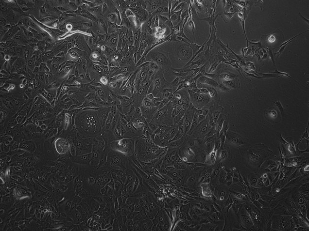
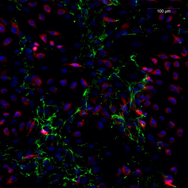
| Phase contrast image of primary tumor cells derived from lung cancer biopsy. | Co-culture of tumor cells derived from a lung biopsy (blue-red: alveolar cells; green: fibroblasts). |
Integration of Cell Culture and Toxicity Assays into microfluidic Chipsystems
The development and creation of applied systems of cytotoxicity determination on different samples and different cell types in vitro requires fast, easy to use and dynamic systems that can provide information about how a sample influences a certain cell type or tissue. A promising way to achieve this is the use of microfluidic whole cell biosensors that combine dynamic and integrated operation modes of microfluidics and complete mammalian (human) cells as complex and stable sensing elements. For the development of these it is important to work on different problems concerning the integration of cells into microfluidic structures. It is preferable to miniaturize the systems and its peripheral equipment and to reduce the technical efforts necessary to run the system. For this the development of incubator independent solutions that need no laminar flow bench for the whole experiment is targeted. Further it is living biological material that shall be supported by the fluidic structures. So the exchange of nutrients and waste products has to be done, stabilization of pH and the space saving generation of stable and physiological temperature conditions have to be implemented. The sterility of the system as well as the stability of the measurement parameters are issues as well as the question how to get the information about the cellular status from the cells without destroying them. To address these problems we developed and tested a chip system that combines the advantages of microfluidic dynamic controlling and operating with highly sensitive senor-cells that can express their physiological status via expression of GFP (Green fluorescent Protein), which can be microscopically detected and quantified. Currently we are working on the integration of primary cell lines into a microfluidic chipsystem. Since these cells are not genetically modified, ECIS-measurements will be used for characterization of cell viability paramters. Future goal is to implement 3D-cell-culture into microfluidic structures for a cell model closer to real conditions.
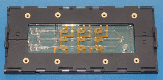
| Lab-on-a-Chip-system for cell culture and toxicity assay. |